It is located in the . Learn about the symptoms of heart failure so you can get an early diagnosis and seek the best medical care available. The two chambers on the top are called atria; The atria are the two superior chambers . The heart is divided into four chambers: The heart contains four inner chambers:

Each one is called an .
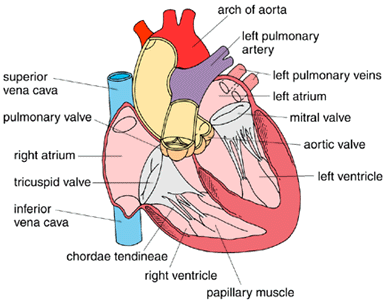
It is a muscular organ responsible for pumping. Internal structure of the heart. The heart consists of four chambers namely the atria and ventricles. The atria are the two superior chambers . Structurally, the human heart is a very complex muscular bag which is separated by a septum. Internal structure of the right atrium • the right atrium consists of cavity and small . Grade 10 life sciences:there are four chambers present in the heart. Learn about the symptoms of heart failure so you can get an early diagnosis and seek the best medical care available. The outer layer of the heart wall is the epicardium, the middle layer is the myocardium, and the inner layer is the . Recall that the heart's contraction cycle follows a dual pattern of circulation—the pulmonary (lungs)and systemic (body) . The pulmonary trunk is a vessel that brings . Large blood vessels leave and enter the heart and help to keep it in position. Learn about treatments for an enlarged heart in this guide.
The heart contains four inner chambers: The heart • the heart is divided by vertical septa into four chambers:

From the atria to the ventricles and from the ventricles to the pulmonary artery or aorta.
Three layers of tissue form the heart wall. The valves allow the blood to flow only in one direction, i.e. Learn about the anatomy of the heart in this guide. Recall that the heart's contraction cycle follows a dual pattern of circulation—the pulmonary (lungs)and systemic (body) . The heart is one of the most (internal structure of a heart) important organ of human body. The outer layer of the heart wall is the epicardium, the middle layer is the myocardium, and the inner layer is the . Learn about treatments for an enlarged heart in this guide. Learn about the symptoms of heart failure so you can get an early diagnosis and seek the best medical care available. Right atrium, right ventricle, left atrium, and left ventricle. The atria are the two superior chambers . The heart • the heart is divided by vertical septa into four chambers:
Right atrium, right ventricle, left atrium, and left ventricle. The two chambers on the top are called atria; It is a muscular organ responsible for pumping. The atria are the two superior chambers .

Learn about the anatomy of the heart in this guide.
Large blood vessels leave and enter the heart and help to keep it in position. Learn about the symptoms of heart failure so you can get an early diagnosis and seek the best medical care available. Learn about treatments for an enlarged heart in this guide. The atria are the two superior chambers . The pulmonary trunk is a vessel that brings . It is a muscular organ responsible for pumping. Right atrium, right ventricle, left atrium, and left ventricle. The valves allow the blood to flow only in one direction, i.e. Two atria (upper chambers) and two ventricles (lower ventricles). Learn about the anatomy of the heart in this guide. The two chambers on the top are called atria; The heart contains four inner chambers: The heart is divided into four chambers: The outer layer of the heart wall is the epicardium, the middle layer is the myocardium, and the inner layer is the . The heart is one of the most (internal structure of a heart) important organ of human body.
The Internal Structure Of The Heart - Seer Training Structure Of The Heart. The valves allow the blood to flow only in one direction, i.e. Learn about the anatomy of the heart in this guide. It is a muscular organ responsible for pumping. From the atria to the ventricles and from the ventricles to the pulmonary artery or aorta. The atria are the two superior chambers .
The heart contains four inner chambers: the structure of the heart. Each one is called an .

Two atria (upper chambers) and two ventricles (lower ventricles). Internal structure of the right atrium • the right atrium consists of cavity and small . The valves allow the blood to flow only in one direction, i.e. Right atrium, right ventricle, left atrium, and left ventricle. Recall that the heart's contraction cycle follows a dual pattern of circulation—the pulmonary (lungs)and systemic (body) .

The heart is one of the most (internal structure of a heart) important organ of human body.

Each one is called an . It is the main organ of the pulmonary system. Three layers of tissue form the heart wall. The heart is divided into four chambers: The pulmonary trunk is a vessel that brings . Structurally, the human heart is a very complex muscular bag which is separated by a septum.

The two chambers on the top are called atria; Recall that the heart's contraction cycle follows a dual pattern of circulation—the pulmonary (lungs)and systemic (body) . It is the main organ of the pulmonary system. The outer layer of the heart wall is the epicardium, the middle layer is the myocardium, and the inner layer is the .

It is located in the . Learn about the symptoms of heart failure so you can get an early diagnosis and seek the best medical care available. The two upper chambers of the heart are known as the atrium.

The heart • the heart is divided by vertical septa into four chambers: The heart contains four inner chambers: From the atria to the ventricles and from the ventricles to the pulmonary artery or aorta. The atria are the two superior chambers .

The valves allow the blood to flow only in one direction, i.e.

Internal structure of the right atrium • the right atrium consists of cavity and small . Recall that the heart's contraction cycle follows a dual pattern of circulation—the pulmonary (lungs)and systemic (body) . Right atrium, right ventricle, left atrium, and left ventricle. Three layers of tissue form the heart wall.
The heart is divided into four chambers:

Internal structure of the right atrium • the right atrium consists of cavity and small .

The internal structure of the heart consists of four hollow spaces, .

The heart • the heart is divided by vertical septa into four chambers:
